Brain Scans in Psychiatry: Transforming Mental Health Care


Intro
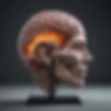
In recent years, the landscape of psychiatry has undergone a notable transformation, with brain scans taking center stage. These imaging technologies such as MRI (Magnetic Resonance Imaging) and PET (Positron Emission Tomography) have progressively shifted from being adjunct tools to playing a pivotal role in diagnosing and managing mental health conditions. The implications of these scans extend beyond mere diagnostics—they offer a window into the complexities of the human mind, challenging traditional methodologies and calling into question established practices.
Recognizing the significance of neuroimaging in psychiatry requires a nuanced understanding of its evolution, current applications, and potential future directions. This examination will delve into the various imaging techniques employed in psychiatric settings, discussing their diagnostic utilities, therapeutic implications, and the ethical dilemmas they may provoke. By fostering an awareness of these elements, we can better appreciate how advances in brain scanning technologies are set to revolutionize psychiatric care.
Research Overview
Summary of Key Findings
A thorough exploration of the literature reveals several key findings that underline the role of brain scans in psychiatric practice:
- Enhanced Diagnostic Accuracy: Neuroimaging has been shown to improve the accuracy of diagnoses in conditions such as schizophrenia, depression, and bipolar disorder, facilitating earlier and more effective interventions.
- Biological Correlates of Mental Illness: Brain scans can illustrate physical changes associated with mental health disorders, helping clinicians and researchers link psychological symptoms with biological abnormalities.
- Treatment Monitoring: Imaging techniques serve as valuable tools for monitoring treatment efficacy and adjusting therapies based on real-time neurobiological feedback.
Relevance to Current Scientific Discussions
The integration of brain scans into psychiatric practice is a hot topic among researchers and clinicians alike. Discussions often revolve around:
- The need for standardized protocols in the application of neuroimaging in clinical settings.
- The potential for personalized medicine, using brain scan data to tailor treatment plans for individuals based on their unique neurobiological profiles.
- Ethical considerations surrounding privacy, consent, and the implications of labeling individuals based on their brain activity.
Methodology
Research Design and Approach
In examining the role of brain scans in psychiatry, a qualitative approach can provide deeper insights into how these tools are currently perceived and utilized in clinical environments. This research involves:
- Literature Review: Analyzing peer-reviewed studies, clinical trials, and expert interviews provides a broad understanding of current knowledge and emerging trends.
- Case Studies: Examining specific instances where neuroimaging significantly influenced psychiatric diagnoses or treatment can illuminate practical applications.
Data Collection and Analysis Techniques
Data collection methods include:
- Gathering primary data through interviews with mental health professionals about their experiences with neuroimaging.
- Collecting secondary data from existing databases and literature to analyze how brain scans have historically been used in psychiatry.
Analysis techniques will involve thematic analysis to identify recurring themes and issues surrounding the application and implications of brain scans in mental health care.
"Neuroimaging is not just about seeing the brain; it’s about understanding its stories."
Through this comprehensive examination, we aim to illuminate the critical interplay between neuroimaging technologies and psychiatric practice, ultimately enriching the dialogue around mental health care.
Preface to Brain Scans in Psychiatry
Brain scans have become an indispensable tool in psychiatry, offering unique insights beyond traditional clinical assessments. They allow for a window into the living brain, providing objective data that can affirm or contradict subjective patient reports. This fusion of modern technology and mental health diagnostics holds the promise of refining our understanding of psychiatric disorders and enhancing treatment methodologies.
Importantly, neuroimaging techniques can illuminate the underlying biological factors contributing to mental health issues. This is especially crucial in an era where the stigma surrounding mental illnesses continues to loom large. By embracing neuroimaging, psychiatry can transition from purely behavioral assessments to evidence-based approaches that incorporate biological insights.
Historical Context
The journey toward utilizing brain scans in mental health began decades ago. In the mid-20th century, the evolution of imaging technologies started to alter the landscape of psychiatry. With the advent of technologies like X-ray and later CT scans, physicians began to see a shift towards examining the physical aspects of the brain. However, these early developments were met with skepticism from some in the field.
By the late 20th century, as imaging techniques became more sophisticated, neuroimaging began gaining respect. The introduction of magnetic resonance imaging (MRI) served as a pivotal moment. For the first time, healthcare professionals could image the brain in a non-invasive manner. This ushered in a new era of research, enabling studies that sought to correlate brain structures with various psychiatric disorders such as schizophrenia and depression.
Today, neuroimaging is regarded as a vital contributor to psychiatric research and practice. Instead of just guessing the underpinnings of mental disorders, psychiatry now has empirical data to formulate treatment plans tailored to individual needs.
Importance of Neuroimaging
The impact of neuroimaging on psychiatry cannot be overstated. By employing various imaging techniques, clinicians can better understand the neurobiological correlates of mental health disorders. For instance, studies have shown that individuals with major depressive disorder often exhibit distinct patterns of activity in specific brain regions.


Neuroimaging also plays a significant role in:
- Diagnosing psychiatric disorders: By revealing abnormalities in brain structure and function, scans can help identify conditions such as bipolar disorder or post-traumatic stress disorder (PTSD).
- Personalizing treatment plans: Understanding brain activity can guide the choice of therapeutic approaches. This is especially salient when dealing with medication resistance, where standard treatments fail to alleviate symptoms.
- Monitoring treatment efficacy: By comparing scans before and after treatment interventions, practitioners can evaluate which therapies yield the best results for individual patients.
Neuroimaging holds potential for both research and clinical practice. As technology continues to advance, the ability to connect mental health disorders with specific neural pathways is likely to grow, paving the way for more targeted and effective treatments. In an age where mental health is finally being acknowledged, brain scans offer a crucial piece of the puzzle.
Overview of Imaging Techniques
The landscape of psychiatric practice is undergoing significant transformation, largely thanks to advancements in neuroimaging techniques. Understanding these imaging methods is crucial, as they serve not only as diagnostic tools but also as gateways to comprehending the complexities of mental health disorders. Each technique possesses its unique strengths and limitations, shedding light on various aspects of brain function and pathology.
In the realm of psychiatry, where the brain's intricate workings often elude simplistic explanations, imaging techniques provide concrete insights into brain activity, structure, and biochemistry. This section delves into four prominent imaging modalities: Magnetic Resonance Imaging (MRI), Positron Emission Tomography (PET), Computed Tomography (CT), and Functional MRI (fMRI). The depth of analysis for each method will help elucidate how these technologies contribute to improved patient outcomes and enhance our understanding of mental illnesses.
Magnetic Resonance Imaging (MRI)
Magnetic Resonance Imaging is a non-invasive imaging technique that allows for detailed visualization of brain anatomy. Unlike X-rays or CT scans, MRI uses strong magnetic fields and radio waves to create images. One of its primary benefits is its high resolution, which makes it particularly useful for examining structural abnormalities such as tumors or lesions. Furthermore, MRI can visualize different types of brain tissue, helping clinicians distinguish between gray and white matter.
Another significant advantage of MRI is its ability to provide insights into chronic conditions. For example, it can assist in diagnosing neurodegenerative diseases like Alzheimer's by identifying characteristic brain atrophy patterns. While MRI is primarily structural, enhancements like diffusion tensor imaging can reveal the integrity of white matter pathways, making it increasingly valuable for assessing psychiatric conditions such as schizophrenia or major depressive disorder.
Positron Emission Tomography (PET)
Positron Emission Tomography stands out by allowing us to observe metabolic processes in the brain. Through the injection of a radiotracer, PET scans measure regional cerebral blood flow and glucose metabolism. This is particularly useful in psychiatric settings, where traditional imaging might not capture functional nuances. With PET, clinicians can assess how different brain regions communicate and work together during specific tasks.
For instance, research has shown altered glucose metabolism in the brains of individuals with depression, providing a biochemical perspective on the disorder that goes beyond mere symptoms. However, it’s worth noting that PET’s limitations include cost and exposure to radiation, which means it should be applied judiciously in psychiatric evaluations.
Computed Tomography (CT)
Computed Tomography, often called a CT scan, offers quick imaging and is readily available in many medical settings. Using X-rays from various angles, it creates cross-sectional images of the brain. While not as detailed as MRI, CT is often the go-to method for initial evaluations, especially in emergency cases, such as assessing head injuries or hemorrhages.
CT scans can also help identify structural issues like tumors or abnormalities that could contribute to psychiatric symptoms. However, reliance on CT alone can lead to oversights, as it may not reveal subtle mental health issues that require more advanced imaging methods. Therefore, in psychiatric contexts, CT is typically considered a complementary approach rather than a standalone diagnostic tool.
Functional MRI (fMRI)
Functional MRI is a game-changer, as it allows clinicians to examine brain activity in real-time. By detecting changes in blood flow associated with neural activity, this technique provides an indirect measure of brain function. fMRI can reveal how different regions of the brain are activated during tasks, which is crucial for understanding cognitive processes involved in mental illnesses.
For example, fMRI has been instrumental in shedding light on how individuals with anxiety disorders process fear-related stimuli versus individuals without such disorders. This information can guide treatment decisions by revealing which therapeutic approaches may be more effective. One of the challenges with fMRI, however, is the interpretation of results, as factors like task design and participant variability can influence outcomes.
In summary, the suite of imaging techniques available in psychiatry provides essential insights into both the structure and function of the brain, significantly aiding diagnosis and treatment planning. Understanding the nuances of each method is vital for optimizing patient care and advancing psychiatric research.
The combination of these imaging modalities enhances our understanding of mental health conditions, pushing forward the boundaries in both research and clinical practice.
Applications in Psychiatric Diagnosis
The field of psychiatry has traditionally relied on interviews, observations, and self-reported symptoms to diagnose mental health disorders. However, as science digs deeper into the mind's complexities, the role of brain scans has become increasingly vital. Understanding how neuroimaging techniques can be applied in psychiatric diagnosis not only enhances clinical accuracy but also opens new pathways in patient care and treatment. This section unpacks the major contributions of brain scans in this context, focusing on their ability to identify psychiatric disorders, differentiate between them, and assess treatment responses.
Identifying Psychiatric Disorders
Brain imaging plays a crucial part in unveiling the latent markers of various psychiatric disorders. Techniques like fMRI can illustrate changes in brain activity patterns, giving professionals insight into conditions that might otherwise remain undetectable. For example, studies have shown that individuals with depression might display altered connectivity in the default mode network, which is pivotal in self-referential thought processes.
With neuroimaging, practitioners can obtain a clearer picture of how a patient’s brain functions, revealing anomalies that correlate with diagnosed conditions such as schizophrenia or bipolar disorder. Thus, scans serve as complementary tools, helping to refine diagnoses that are often subjective and prone to error.
Differentiating Between Disorders
One of the standout advantages of incorporating brain scans into psychiatric assessments is the ability to differentiate between disorders that share similar symptoms. For instance, anxiety and depression can present overlapping characteristics, complicating diagnosis. However, neuroimaging studies have indicated differential brain patterns in these disorders. Using a PET scan, clinicians might observe variations in metabolic activity between a patient with anxiety versus one with a major depressive disorder.
This differentiation is critical. Misdiagnosis not only impacts treatment efficacy but often leads to severe consequences for the patient's quality of life. By leveraging brain scans, practitioners can navigate the murky waters of psychiatric diagnostics with greater confidence.
Assessment of Treatment Responses
Measuring treatment efficacy is another area where brain scans shine. They not only reflect how a patient's brain responds to medications but also gauge the impact of therapeutic interventions. For example, follow-up fMRI scans might reveal decreased activity in certain brain regions following cognitive-behavioral therapy for PTSD, indicating that the treatment is successfully modifying the patient's neural pathways.


Moreover, assessing treatment responses through neuroimaging can identify when a patient is not responding as expected. This allows clinicians to make timely adjustments to treatment plans, optimizing outcomes. The adaptability afforded by imaging techniques empowers practitioners to create more tailored treatment strategies, potentially enhancing recovery rates.
"Neuroimaging could well be the compass we need in the complex terrain of psychiatric disorders. It guides us, highlighting paths we would otherwise overlook."
Overall, the applications of brain scans in psychiatric diagnosis represent a significant leap forward in mental health care. By integrating these innovative technologies into clinical practice, providers can better identify disorders, differentiate between them, and evaluate treatment responses effectively. This not only benefits individual patients but also enhances the collective understanding of mental health disorders in the broader context of psychological research.
Research Developments in Neuroimaging
Research developments in neuroimaging have become an underlying force in reshaping the landscape of psychiatric diagnosis and treatment. As technology evolves, so does our ability to understand the brain’s intricate workings, especially in the context of mental health. This section delves into pivotal strides made in the field, showcasing recent studies, innovative imaging technologies, and the integration of genetic data, all of which hold promise for enhancing psychiatric practice.
Recent Studies and Findings
Numerous studies have emerged that significantly advance our grasp of the relationship between neuroimaging and psychiatric disorders. For instance, investigations into the neural correlates of depression have shown that abnormalities in brain areas like the prefrontal cortex and amygdala are common. These findings have been grounded in substantial brain scan data, such as fMRI studies that reveal altered activity patterns when patients perceive emotional stimuli.
Moreover, a recent meta-analysis compiled results from multiple studies focusing on schizophrenia. This analysis highlighted a distinct pattern of volumetric changes in specific brain regions among affected individuals compared to healthy controls. Such consistent findings across varied populations not only strengthen the validity of imaging as a diagnostic tool but also push the boundaries of personalized treatment options.
"Neuroimaging allows us to visualize the brain and gives tangible evidence to what was previously considered only subjective experience."
Advancements in Imaging Technology
The technological landscape surrounding neuroimaging constantly improves, enabling more precise and detailed scans. Innovations such as high-field MRI scanners enhance resolution and sensitivity. These upgrades allow researchers and clinicians to evaluate fine details like microstructural changes in the white matter, shedding light on conditions like multiple sclerosis or autism.
Developments in diffusion tensor imaging (DTI) have further broadened our understanding by mapping the brain’s white matter tracts effectively. This technique helps in understanding how different brain regions communicate and can provide insights into disorders characterized by disrupted connectivity, such as bipolar disorder.
Importantly, the advent of machine learning algorithms applied to neuroimaging data is revolutionizing the way we interpret complex patterns. By utilizing extensive datasets, these algorithms can predict diagnoses with remarkable accuracy, paving the way for innovative diagnostic methods.
Integrating Genetic Data with Imaging
The convergence of neuroimaging and genetic research introduces a compelling frontier in mental health studies. By correlating specific genetic markers with neuroimaging results, researchers can begin to unravel the biological underpinnings of psychiatric disorders. For example, studies investigating the genes associated with serotonin regulation have correlated genetic variations with altered brain function observable in SPECT and PET scans.
This integration helps stratify patient populations, allowing for a deeper understanding of the mechanisms at play and enhancing the development of targeted therapies. Integrating genotyping with neuroimaging could lead to breakthroughs in personalized medicine, where treatment plans align more closely with each individual’s unique biological makeup.
In summary, the ongoing research developments in neuroimaging not only bolster our understanding of psychiatric disorders but also offer pathways towards refining diagnostics and tailoring treatments that are quicker and more effective. As both imaging technologies and genetic insights evolve, the future holds considerable promise for advancing psychiatric care.
Ethical Considerations in Brain Scanning
The incorporation of brain scans into psychiatric practice raises a multitude of ethical questions that cannot be overlooked. As we dive deeper into the interplay between technology and mental health, understanding these ethical dimensions becomes crucial. Ethics in brain scanning isn't just a checklist. It's about how sensitive data is handled, the ramifications of misinterpretation, and the potential societal impacts that could stem from advances in neuroimaging. Every scan taken holds not just images but potentially profound implications for patients’ lives and the mental health field as a whole.
Patient Privacy and Consent
In the realm of psychiatry, patient privacy is sacrosanct. Brain scans can reveal intimate details about a person's mental state, possibly more than any verbal disclosure during therapy. As technology evolves, so do the techniques to extract data from these scans. Therefore, robust measures must be in place to ensure that such data remains confidential. Not to mention, there is a matter of informed consent. Patients must fully understand what it means to undergo a brain scan: the procedure itself, what the results might reveal, and how that information could be used. The discussion regarding the scope of their consent – in or out of the clinical setting – is vital.
"Informed consent must be a dynamic conversation, not a one-time form to sign."
When patients feel a sense of agency over their health data, it fosters trust. This trust is paramount for an effective therapeutic alliance. With the capability to analyze brain function comes the responsibility to handle that information judiciously, respecting how it may affect a patient's life and relationships.
Implications of Misinterpretation
The interpretation of brain scans is not foolproof. These images can be nuanced; small variations may lead to different conclusions. False positives or negatives could lead to misdiagnoses, resulting in inappropriate treatment plans. For instance, if a scan indicates a possible malfunction in brain function related to anxiety but is wrongly interpreted as a sign of schizophrenia, the implications for treatment decisions can be drastic.
Moreover, public perception adds another layer of complexity. If results from neuroimaging are misrepresented or sensationalized in media outlets, it can skew public understanding and fuel stigma around mental health issues. The potential for misinterpretation also raises ethical concerns about who has the authority to read these scans and the standards by which they evaluate them. Therefore, a rigorous and standardized training for professionals interpreting these images is imperative.
The interplay of brain imaging, ethics, and psychiatric care necessitates ongoing conversation and vigilance. As neuroimaging technology becomes more integrated into psychiatric practice, stakeholders must continually negotiate the ethical landscape to protect individuals while harnessing the potential benefits. Understanding these considerations ensures that we tread carefully on the delicate grounds where science meets humanity.
Challenges and Limitations
The field of psychiatry, while evolving rapidly thanks to the rise of neuroimaging, is not without its hurdles. Understanding these challenges and limitations is crucial for both professionals in the field and those seeking to benefit from it. The integration of brain scans into psychiatric practice can provide remarkable insights, yet pitfalls remain that can undermine the effectiveness of these sophisticated tools.
Navigating through the advancements and maintaining a mindful posture towards the limitations has become a significant consideration for researchers and clinicians alike. The overarching aim should always be to better serve patients while ensuring the integrity of the diagnostic processes.


Technical Limitations of Scanning Techniques
Even though neuroimaging techniques like MRI and PET scans have made significant strides, they come with technical limitations that can affect their utility. Firstly, the cost associated with these advanced imaging modalities can be prohibitive for some institutions and patients. The machinery required is often expensive, and the need for specialized training adds another layer of financial burden.
Additionally, each scanning technique has its biases. For example, Magnetic Resonance Imaging (MRI) is excellent for structural imaging but falls short in exploring metabolic functions of the brain. In contrast, Positron Emission Tomography (PET) provides metabolic data but lacks the spatial resolution that MRI offers. This can lead to scenarios where reliance on one technique overshadows the necessity of a multi-faceted diagnostic approach.
Another technical barrier is motion artifact. Patients, especially those experiencing severe psychiatric symptoms, may have difficulty staying still for extended periods during scans, leading to compromised image quality. Researchers must thus take this into account when interpreting results, recognizing that motion can artificially alter findings, preventing accurate clinical conclusions.
Variability in Imaging Results
Variability in imaging results poses a critical challenge in establishing a standard for psychiatric diagnosis. Even when scans are executed properly, variances in brain anatomy and function can yield differing interpretations. What may appear as a sign of a specific disorder in one individual might not manifest in another, even if both experience similar symptoms.
This inherent variability challenges the notion of universality in diagnostic imaging. Many factors influence brain scans, including age, genetics, and comorbid conditions. The subjectivity in interpretation further complicates matters. Not all radiologists or psychiatrists may agree on what constitutes a normal or abnormal finding. Therefore, establishing robust guidelines and criteria becomes imperative but remains a formidable task.
Moreover, although many studies contribute valuable insights into imaging patterns associated with specific psychiatric conditions, generalizing findings can be problematic. The existing database of healthy versus disordered brains is limited and often skewed towards certain demographics, which can lead to biased interpretations. If researchers can’t agree on the reliability of data, how can clinicians make informed decisions?
The integration of neuroimaging in psychiatry is a double-edged sword; it holds great promise but also carries substantial responsibility.
Thus, while the advantages of integrating imaging into psychiatric practice are clear, acknowledging and addressing these technical and interpretive challenges is essential for progress. As researchers continue to refine methods and develop clearer correlations between brain activity and mental health disorders, clinicians must remain cautious and informed about the limitations as they endeavor to apply neuroimaging to their practice.
Future Directions in Psychiatric Imaging
The landscape of psychiatric imaging is on the brink of a transformative shift. With advancements in technology and an increasing understanding of the brain's complexities, future directions in this field are set to redefine both diagnosis and treatment in psychiatry. As society evolves, so does the demand for more precise, personalized care in mental health, an area where imaging plays a pivotal role. Here’s a close look at emerging technologies and collaborative research initiatives shaping this future.
Emerging Technologies
As we peer into the horizon, several emerging technologies in imaging are eliciting significant interest. One notable advancement is the rise of machine learning algorithms. These algorithms can analyze vast amounts of imaging data, identifying patterns that may be invisible to the human eye. By distinguishing subtle differences in brain activity linked to various psychiatric disorders, machine learning holds promise for enhancing diagnostic accuracy.
Another notable innovation is multimodal neuroimaging. This technique integrates different types of scans, such as fMRI and PET, to create a more comprehensive view of brain function. This holistic approach can provide deeper insights into complex disorders like schizophrenia and bipolar disorder. It allows clinicians to visualize both structure and activity, fostering a more robust understanding of how various disorders manifest.
Telemedicine in psychiatric consultations also takes a front seat. The advent of remote imaging capabilities grants patients more access to diagnostic tools. This not only increases convenience but may also help bridge gaps in underserved areas, ensuring that mental health care reaches a wider population.
"The integration of AI and advanced imaging reshapes the future, promising not merely better diagnostics but also personalized treatment pathways that cater to individual patient needs."
Collaborative Research Initiatives
The drive towards improved psychiatric imaging is bolstered significantly by collaborative research initiatives. In this field, cooperation between universities, healthcare institutions, and tech companies can yield unprecedented results. For instance, partnerships that leverage shared databases of imaging studies can promote large-scale analyses. These analyses lead to the discovery of biomarkers for mental health disorders, streamlining the diagnostic process and treatment planning.
Additionally, funding bodies increasingly prioritize cross-disciplinary projects that blend psychology, neuroscience, and engineering. Such initiatives enrich the data pool and enhance methodological rigor, paving the way for innovations that tackle the nuances of psychiatric conditions.
Moreover, patient involvement in research has gained traction. Engaging patients not only cultivates trust but also ensures that studies reflect real-world applications. Their experiences can steer research focus toward practical challenges, thereby creating solutions that are both relevant and impactful.
Ultimately, as psychiatric imaging moves forward, it will likely capitalize on these emerging technologies and collaborative efforts. By turning promise into practice, the future holds the potential to foster a more precise, compassionate, and effective approach to mental health care.
Finale: The Evolving Role of Neuroimaging in Psychiatry
The contribution of neuroimaging to psychiatry is no longer a mere afterthought, but a cornerstone of modern mental health practice. It brings clarity to murky waters of psychiatric diagnosis, which often rely on subjective assessments. Brain scans, such as MRI and fMRI, allow clinicians to visualize brain activity and structure, shedding light on the biological aspects underlying mental disorders. This is crucial because understanding the physiological basis of conditions—such as depression, anxiety disorders, or schizophrenia—can profoundly affect treatment approaches.
Neuroimaging isn't just a tool; it's reshaping the mental health landscape. By generating data-driven insights, these technologies lead to more informed clinical decisions. For instance, identifying anomalies in brain connectivity can offer vital clues for tailoring therapies that are personalized, thereby increasing the likelihood of successful outcomes.
Summary of Insights
As we’ve traversed the research and applications of neuroimaging in psychiatry, several key insights emerge:
- Enhanced Diagnostics: Brain imaging significantly improves diagnostic accuracy, allowing for differentiation between disorders that might otherwise appear similar based on symptoms alone.
- Treatment Monitoring: Scans not only assist in identifying issues but also in monitoring the efficacy of treatment over time, thus facilitating adjustments based on real-time data.
- Research Advancements: Current advancements underscore a vibrant field where findings can spark new hypotheses, enhancing our overall understanding of mental illnesses.
It’s important to remember, however, that the advent of neuroimaging does not diminish the value of traditional assessment methods but rather complements them. This dual approach harmonizes the art and science of mental health care, where subjective experiences meet objective data.
The Path Forward
Looking ahead, the role of neuroimaging in psychiatric settings will likely expand even further as technology and methodologies improve. Here are some areas to watch:
- Integration with Other Modalities: Combining neuroimaging data with genetic information, psychological evaluations, and patient history can lead to a more holistic understanding of mental health.
- Technological Innovations: Emerging technologies, like machine learning algorithms applied to imaging data, might help predict treatment responses or the likelihood of developing certain disorders.
- Global Partnerships: Collaborative efforts between research institutions and clinical settings can democratize the benefits of neuroimaging, ensuring diverse populations receive equitable access to these innovations.
In summary, embracing neuroimaging as a vital resource in psychiatry enriches both diagnosis and treatment, propelling mental health care into a new era where insights are gleaned from the very structure and function of the brain itself. Future developments promise to deepen our grasp of mental health, transforming how we approach not only treatment but also understanding the profound complexities of the human mind.


